
Source: www.realites-ophtalmologiques.com
by O. Sandalion July 14, 2015 in General Reviews
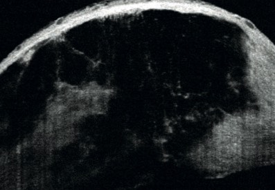 The improvement in resolution of the latest generation OCT allows for precise evaluation of the keratoconic cornea. Thanks to the study of epithelial mapping, OCT enhances the detection of subclinical keratoconus.
By studying the changes in corneal structure and precisely measuring the thickness of the different corneal layers, OCT has become an essential tool for optimal management of keratoconus.
The improvement in resolution of the latest generation OCT allows for precise evaluation of the keratoconic cornea. Thanks to the study of epithelial mapping, OCT enhances the detection of subclinical keratoconus.
By studying the changes in corneal structure and precisely measuring the thickness of the different corneal layers, OCT has become an essential tool for optimal management of keratoconus.Contents
1. Diagnosis of subclinical keratoconus
2. Precise anatomical diagnosis of keratoconus of varying severity and OCT classification of keratoconus
Keratoconus is a corneal dystrophy manifested by a non-inflammatory protrusion of the cornea, with a slowly progressive and irreversible course, generally appearing during adolescence. The disease rarely progresses after the age of 40. This disease is characterized by thinning of the corneal stroma, a decrease in the number of collagen fibrils, keratocyte apoptosis, and abnormalities in Bowman’s membrane [1]. To date, the diagnosis and management of keratoconus relies mainly on corneal topography thanks to the measurement of the degree of corneal deformation (keratometry) and the approximate assessment of corneal thickness (pachymetry) [2]. These two criteria are certainly important, but they do not take into account the anatomical changes that occur during the various stages of keratoconus progression. The improvement in resolution of recent OCTs, spectral domain and Fourier domain, allows for an accurate study of corneal architecture, improving the screening of subclinical keratoconus and the precise diagnosis of the severity of established keratoconus [3].
Diagnosis of subclinical keratoconus
The diagnosis of confirmed forms is generally easy with topographic examination. On the other hand, screening for early forms (subclinical or subtlest forms) remains more problematic and a real challenge, especially in refractive surgery.
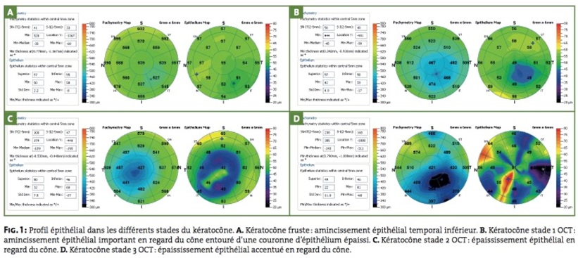
Keratoconus is accompanied by early changes at the level of the epithelium, which, due to its "smoothing" role, restores the regularity of the corneal surface and can mask subclinical forms (fig. 1). The study of the epithelial profile by OCT thus provides important elements for the diagnosis of subtle forms.
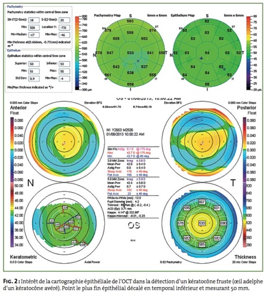
In a recent study of 36 patients with subclinical keratoconus (fellow eyes of a confirmed keratoconus and with normal topography: negative Orbscan indices (K ≤ 47D and I-S ≤ 1.4) and Pentacam (KI < 1.03, KCI < 1.07) and TKC, we showed that an inferior location of the thinnest epithelial point as well as a thickness of this point less than 52 μm were suspicious for subclinical keratoconus [4] (fig. 2). The ‘doughnut’ or ‘bagel’ appearance of the epithelium, initially described by Reinstein, is also highly suggestive of keratoconus: localized epithelial thinning at the apex surrounded by a crown of thickened epithelium [5]. The OCT pachymetric map can also show an inferotemporal displacement of the thinnest point.
The analysis of the different corneal layers at this stage shows no abnormalities.
Precise anatomical diagnosis of variably severe keratoconus and OCT classification of keratoconus
Corneal topography allows diagnosis from the early stages, but does not allow precise assessment of the anatomical changes of the keratoconic cornea. The OCT pachymetric map is useful for the diagnosis of keratoconus by showing an inferoparacentral corneal thinning and a difference greater than 45 μm between superior nasal pachymetry and inferior temporal pachymetry over 5 mm of the keratoconic cornea [6].
Recently, in a study of 218 keratoconic patients, we established a new anatomical classification based on OCT (Optovue with 6 μm resolution) [7], which includes five stages (fig. 3):
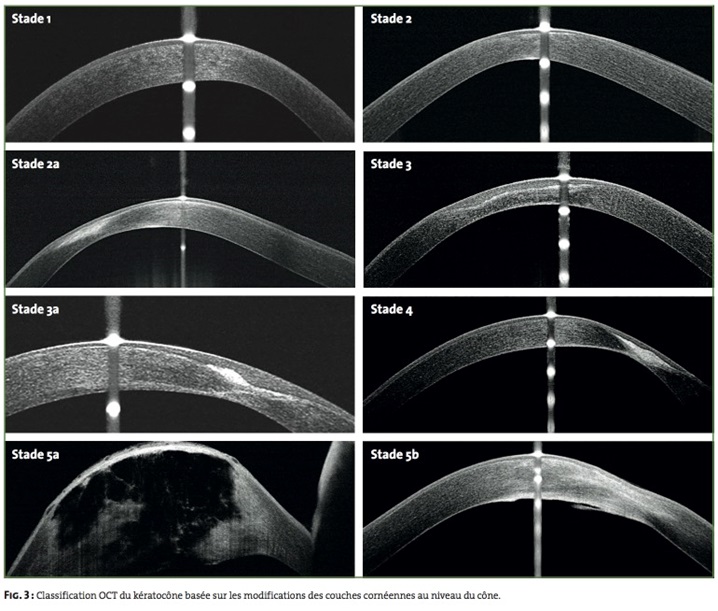
- stage 1: thinning of the epithelial and stromal layers of normal appearance at the corneal cone. In a recent study, Yadav, using a prototype 1.1 μm OCT, showed that there is thinning of Bowman’s membrane thickness [8];
- stage 2: hyperreflectivity at the level of Bowman’s layer and epithelial thickening facing the cone;
- stage 3: posterior invagination of hyperreflective structures at Bowman’s layer, with increased epithelial thickening and stromal thinning;
- stage 4: panstromal scar at the cone;
- stage 5: hydrops. Acute stage 5a: rupture of Descemet’s membrane with severe corneal edema, tearing of collagen lamellae and formation of intrastromal and intraepithelial cysts. Scarred stage 5b: panstromal scar and residual aspect of Descemet’s membrane rupture.
Vogt striae were observed in various OCT stages (stages 1, 2 and 3) in our series, and were not considered as criteria for classification. They appear as hyporeflective stromal bands between Descemet’s membrane and Bowman’s layer and not as folds of Descemet’s membrane as previously suggested.
The study of the OCT pachymetric map is essential before performing cross-linking, keratoplasty, or placement of an intrastromal ring.
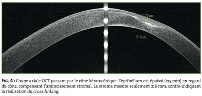
OCT pachymetry is more precise than that of current topographers for the measurement of minimum pachymetry (cross-linking) and at 5-6 mm (placement of intrastromal rings). From stage 2 of the OCT classification of keratoconus, epithelial thickening can mask stromal thinning if only total pachymetry is considered. A minimum pachymetry of 400 μm, for example, may correspond to 70 μm of epithelial thickness and 330 μm of stromal thickness, contraindicating classic cross-linking, whereas it is not if only the total pachymetry measured by corneal topography or ultrasound pachymetry is considered (fig. 4).
The results of the study of the integrity of the Descemet membrane (scarring from ruptures) and the location of the stromal opacities in relation to it are important to know before performing a deep lamellar graft.
Bibliography
1. Amsler M. Classic keratoconus and subclinical keratoconus; unitary arguments. Ophthalmologica, 1946;111:96-101.
2.Li X, Yang H, Rabinowitz YS. Keratoconus: classification scheme based on videokeratography and clinical signs. J Cataract Refract Surg, 2009;35:1597-1603.
3.Li Y, Tan O, Brass R et al. Corneal epithelial thickness mapping by Fourier-domain optical coherence tomography in normal and keratoconic eyes. Ophthalmology, 2012;119:2425-2433.
4. Temstet C, Sandali O, Bouheraoua N et al. Corneal epithelial thickness mapping by Fourier-domain optical coherence tomography for detection of subclinical keratoconus. J Cataract Refract Surg, 2014 (in press).
5.Reinstein DZ, Archer TJ, Gobbe M. Corneal epithelial thickness profile in the diagnosis of keratoconus. J Refract Surg, 2009;25:604-610.
6.Li Y1, Meisler DM, Tang M et al. Keratoconus diagnosis with optical coherence tomography pachymetry mapping. Ophthalmology, 2008;115:2159-2166.
7. Sandali O, El Sanharawi M, Temstet C et al. Fourier-Domain optical coherence tomography imaging in keratoconus: a corneal structural classification. Ophthalmology, 2013;120:2403-2412.
8. Yadav R, Kottaiyan R, Ahmad K et al. Epithelium and Bowman’s layer thickness and light scatter in keratoconic cornea evaluated using ultrahigh resolution optical coherence tomography. J Biomed Opt, 2012;17:116010.The authors have declared that they have no conflicts of interest regarding the data published in this article.